Tympanometry is a critical tool for diagnosing and evaluating middle ear function. In this article, we will explore what tympanometry is, break down the various types of tympanograms, guide you on how to interpret tympanometry test results, and clarify the significant distinctions between clinical and portable tympanometers.
What is Tympanometry?
Tympanometry is a non-invasive audiological test used to evaluate the health and functionality of the middle ear. It provides valuable insights into the condition of the eardrum (tympanic membrane) and the middle ear, specifically the mobility of the ossicular chain (tiny bones in the middle ear) and the presence of any fluid, eardrum perforations or blockages.
Tympanometry involves the application of both negative and positive pressure to the middle ear while maintaining a constant probe tone. Through tympanometry, hearing care professionals evaluate the absorption and reflection of the probe tone within the middle and inner ear. This assessment enables clinicians to gain insights into the behavior and functionality of the middle ear system (tympanic membrane, ossicular chain and eustachian tube).
What is a Tympanometry Test?
A tympanometry test is a non-invasive assessment of the compliance of the middle ear, i.e., its ability to transmit sound effectively. It measures the impedance of the middle ear by varying the air pressure within the ear canal and recording the corresponding changes in the eardrum's movement.
The tester inserts a small probe into the ear canal during a typical tympanometry test. The probe consists of a speaker and a microphone (and an air pressure tube). The speaker emits a pure tone sound, which travels into the ear canal, while the microphone measures the sound reflected back. The air pressure tube is connected to a pump that systematically varies the air pressure in the ear canal, recording the amount of reflected sound as the air pressure changes.
What is a Tympanogram?
A tympanogram is a graphical representation generated through tympanometry, typically displaying the motion of the tympanic membrane. It commonly takes on a pyramid-like shape. The highest point on a tympanogram corresponds to when the eardrum experiences equilibrium between negative and positive pressure. With a classification framework like Jerger's template, it becomes feasible to ascertain whether the tympanic membrane exhibits unrestricted movement or if there is an underlying disorder.
How to Read a Tympanometry Test
Tympanometry results are typically represented on a graph, known as a tympanogram. The chart displays compliance (y-axis) to air pressure (x-axis). By analyzing the shape and position of the tympanogram, hearing care professionals can make informed assessments about the middle ear's functionality. Here are the primary tympanometry types, as purposed by Jerger:
Tympanometry Type A:
-
This is the ideal tympanogram and indicates normal middle ear function.
-
The graph typically shows a peak when the eardrum has its highest compliance.
-
Type A tympanograms suggest sound is conducted efficiently through the middle ear.

Figure 1. Tympanogram Type A
Tympanometry Type As:
-
Represents a shallow peak on the tympanogram.
-
Indicates reduced compliance in the middle ear system.
-
Often associated with stiffness or reduced mobility of the ossicles, possibly due to conditions, like otosclerosis.
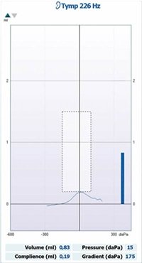
Figure 2. Tympanogram Type As
Tympanometry Type Ad:
-
Features a deeper, more exaggerated peak on the tympanogram.
-
Suggests increased compliance in the middle ear system.
-
Typically associated with flaccid eardrum or ossicular discontinuity, possibly caused by trauma or other ear-related issues.
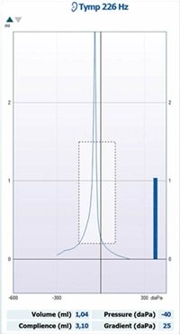
Figure 3. Tympanogram Type Ad
Tympanometry Type B:
-
A Type B tympanogram displays a flat line, indicating little or no compliance in the middle ear.
-
This may suggest issues like a perforated eardrum, fluid in the middle ear, or problems with the ossicles.
-
Further examination is required to identify the specific problem.
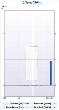
Figure 4. Tympanogram Type B
Tympanometry Type C:
-
Type C tympanograms show a peak, but it occurs when air pressure is more negative than in Type A tympanograms.
-
This suggests negative pressure in the middle ear, which could indicate Eustachian tube dysfunction or the initial or resolving stages of an ear infection.
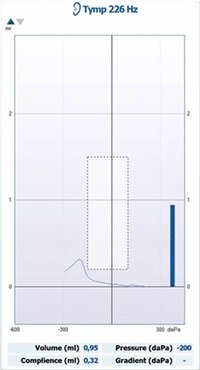
Figure 5. Tympanogram Type C
Tympanometry for Ear drum Perforation:
-
Typically shows a flat or abnormally low peak on the tympanometry graph.
-
The equivalent ear canal volume measurement is larger than normal.
-
Indicates little to no compliance in the middle ear due to the hole or perforation in the eardrum.
-
Suggests that sound is not being effectively transmitted through the middle ear, requiring further examination and appropriate treatment.
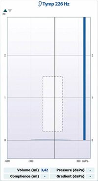
Figure 6. Tympanogram for Ear Drum Perforation
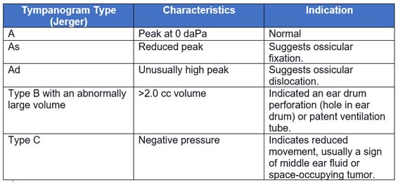
Figure 7. Tympanogram types as purposed by Jerger
What is a Tympanometer?
Tympanometry testing involves using a tympanometer. To conduct the test, a probe is carefully placed into the ear canal, ensuring an airtight seal. Subsequently, the probe administers varying levels of air pressure within the ear canal while simultaneously tracking alterations in the impedance of the middle ear. The collected data is then charted on a tympanogram graph, serving as a valuable source of information for audiologists.
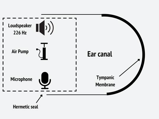
Figure 8: The basic components of a tympanometer. The loudspeaker introduces a 226-Hz tone to the ear canal while the pump varies the air pressure above and below atmospheric pressure in the ear canal. The microphone records the level of the tone at various ear canal pressures.
Clinical Tympanometers versus Portable Tympanometers
Tympanometers come in two primary forms: clinical/ diagnostic and portable/ screening. Each has its own set of advantages and applications:
1. Clinical Tympanometer:
-
Often referred to as "Middle Ear Analyzers" or "Clinical Immittance Systems."
-
Widely used in audiology and ENT clinics, university training programs, and hospital settings.
-
These devices provide a wide range of customizable testing options, making them perfect for comprehensive assessments and diagnosing various ear conditions.
-
Users can opt for either manual or automatic testing procedures.
-
Offer advanced testing protocols, including assessments for eustachian tube function, acoustic reflex decay, acoustic reflex latency, and wide band tympanometry.
Clinical Tympanometer Examples:
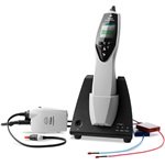
Grason-Stadler TympStar Pro™
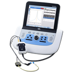
Interacoustics Titan
2. Portable Tympanometer:
-
Portable/Screening tympanometers are typically compact and lightweight, with some designed to be handheld.
-
They are created for the swift and automatic initiation of tympanogram tests once the probe is correctly inserted into the ear canal.
-
Many of these devices offer the option for basic acoustic reflex testing.
-
Some models can be equipped with a screening audiometer function.
-
Minimal training is needed to operate these devices effectively.
-
They serve as an excellent solution for pediatricians, school nurses, or any specialty in need of a quick and straightforward method to screen for middle ear dysfunction.
Screening Tympanometer Examples:
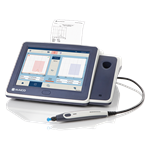
MAICO touchTymp MI 24
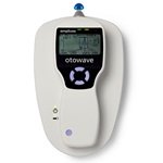
Amplivox Otowave 102 | Portable Tympanometer | e3 Diagnostics
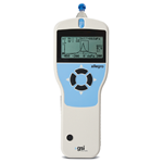
Grason-Stadler Allegro | GSI Tympanometer | e3 Diagnostics
In summary, tympanometry is a vital tool in diagnosing and assessing middle ear function. Understanding the distinct types of tympanometry, reading tympanometry test results, and distinguishing between clinical and portable tympanometers can help hearing care professionals effectively monitor and manage ear health.
Are you considering purchasing a clinical or screening tympanometer? Explore our wide selection here.
Other Good Reads: The Benefits of Wide Band Tympanometry
Follow us on Social!


If you haven't already, make sure to subscribe to our newsletter to keep up-to-date with our latest resources and product information.